

[Author Disclosure: This content reflects the views of the author and is provided for informational purposes only. It does not constitute investment advice and is not a recommendation or endorsement by BioTech Funding Portal. Any investment decision should be based solely on the issuer’s official offering materials.]
By Chris Hopkins, CEO of Glafabra Therapeutics. The author is an employee of Glafabra Therapeutics, which is developing an investigational cell-based therapy for Fabry disease.
Fabry disease is caused by loss of a single lysosomal enzyme, alpha-galactosidase A (α-Gal A), the result of an inherited pathogenic variant in the GLA gene. The absence of this enzyme leads the body to having no way to remove a fatty molecule it produces every day. This fatty lipid builds up to toxic levels over time.
Three facts about Fabry:
- The enzyme deficiency is alpha-galactosidase A.
- The lipid molecule that builds up is globotriaosylceramide.
- The GLA gene sits on the X chromosome so it has sex-specific presentations.
From those three facts, almost everything about the disease follows: who gets it, how they get it, which organs fail, and, more recently, how each class of therapy tries to intervene. The remainder of this article is a primer on what happens to the body from the deficiency, and what the tissues actually look like before and after the treatments that are now available.
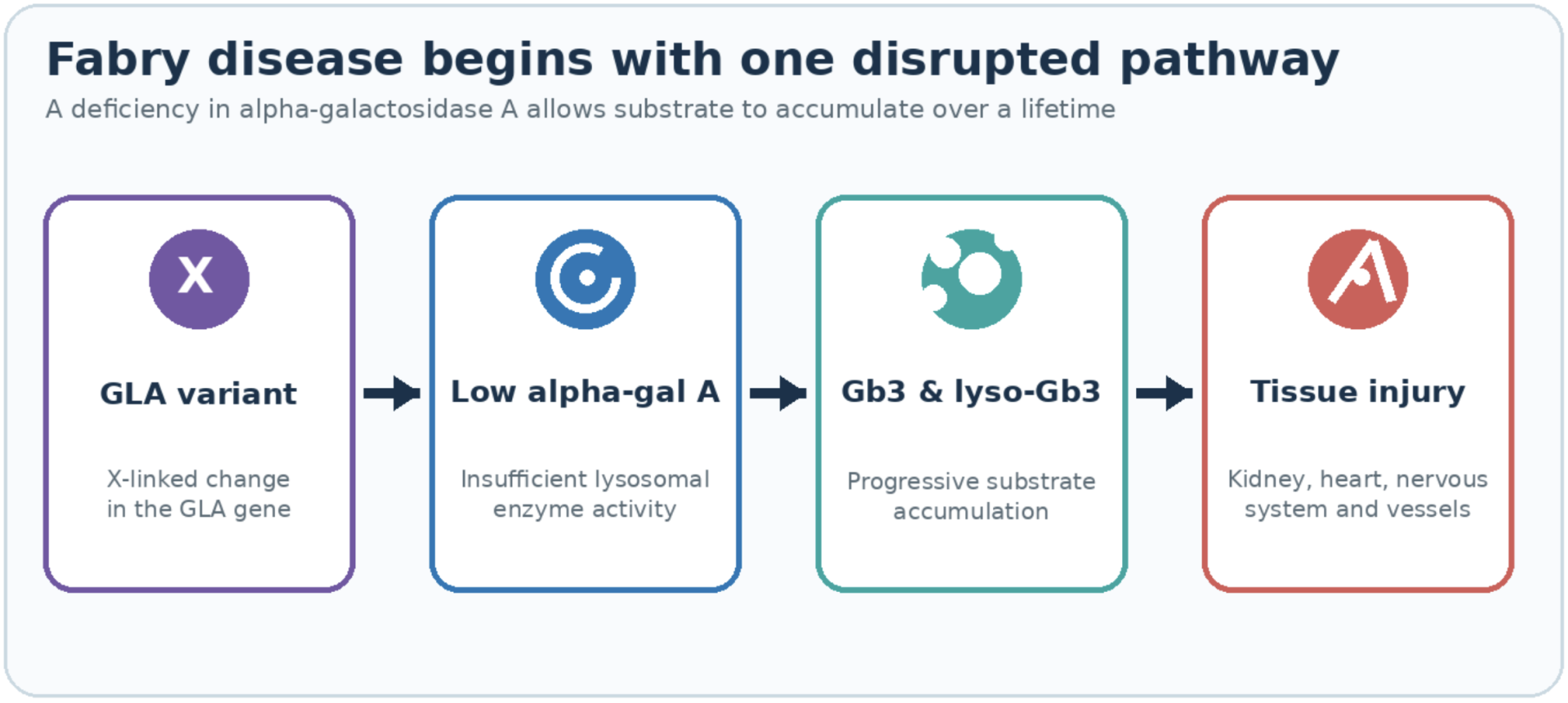
Figure 1. The core Fabry disease cascade: GLA deficiency leads to progressive substrate accumulation and multisystem tissue injury.
The molecular defect
The GLA gene encodes alpha-galactosidase A, a lysosomal hydrolase whose job is to start the breakdown of globotriaosylceramide lipid, which is abbreviated Gb3 or GL-3, by cleaving off a galactose sugar from the end of the fatty lipid. When GLA is mutated and enzyme activity falls, Gb3 can no longer enter its breakdown pathway. Instead, it accumulates inside the lysosomes of cells throughout the body, year after year, starting before birth and continuing for a lifetime.
Gb3 build up is not the whole story. As it starts to build up a second metabolite starts to form: a deacylated, water-soluble derivative called globotriaosylsphingosine, or lyso-Gb3. This molecule readily circulates in plasma and is thought to be one of the main drivers of cellular toxicity (PMID: 37145097). Lyso-Gb3 is elevated in untreated patients, tracks with disease burden, and falls when substrate load is reduced. So, lyso-Gb3 matters as both a diagnostic signal and, increasingly, as a plausible main driver of the tissue injury seen in Fabry disease. When you see a Fabry program report a percentage reduction in lyso-Gb3, that is the number standing in for whether or not the underlying substrate problem is being controlled.
Gb3 accumulation is not confined to one cell type. Vascular endothelium, kidney podocytes and tubular cells, cardiomyocytes, dorsal root ganglion neurons, and smooth muscle all store Gb3. That breadth is why Fabry is a multisystem disease and not an organ-specific one, and it is the central problem any therapy has to solve: reaching every compartment where the substrate collects.
Why the X chromosome changes everything
Because GLA is on the X chromosome, inheritance is X-linked, and this shapes the clinical picture in a way that is frequently misunderstood.
Hemizygous males have a single X and therefore a single copy of GLA. If that copy carries a classic disease-causing mutation, the man has essentially no functional enzyme, and he develops the full, severe, multisystem form. Symptoms in males with the classic phenotype typically begin in childhood or adolescence.
Heterozygous females are not simply "carriers," and treating them as unaffected is a clinical error. Because of random X-chromosome inactivation, each cell in a heterozygous woman expresses either the normal or the mutant GLA allele, and the mosaic that results varies from tissue to tissue and from patient to patient. Some women are nearly asymptomatic; many develop significant cardiac, renal, or cerebrovascular disease, sometimes decades later than affected men, sometimes not much later at all. The range is wide precisely because the degree and pattern of X-inactivation is not uniform. The practical consequence is that female relatives of a diagnosed patient need real evaluation, not reassurance.
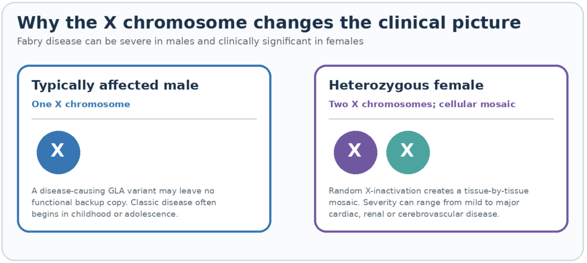
Figure 2. X-linked inheritance produces different—and highly variable—clinical presentations in males and females.
Classic versus later-onset disease
Two broad phenotypes are worth separating because they change who presents, when, and to which specialist.
The classic phenotype arises from mutations that abolish enzyme activity. These patients accumulate Gb3 from early life and show the full syndrome: neuropathic pain, skin lesions, corneal changes, and, over time, progressive kidney, heart, and cerebrovascular disease.
Later-onset variants, often missense mutations that leave some residual enzyme activity, produce a narrower and later picture. Many of these patients come to attention through a single dominant problem, most commonly cardiac, in mid-to-late adulthood. So-called cardiac-variant Fabry is now recognized as an under-diagnosed contributor to unexplained left ventricular hypertrophy, and renal-predominant presentations exist as well. The lesson from the later-onset group is that Fabry is far more common than historical estimates suggested, because for years the milder presentations were simply not being connected to the underlying enzyme deficiency.
What Fabry damages, untreated
Left alone, Fabry is a slow, cumulative disease, and the natural history is the baseline against which every therapy should be read. For instance, classic measures of kidney dysfunction such as estimated glomerular filtration rate (eGFR) can vary widely between Fabry-affected individuals (+/- 2x or more), while a given patient’s day to day variation in eGFR is much smaller. So a good historical data on eGFR decline rate in a patient can be a key biomarker reference point to measure the small slope changes of drug-induced stabilization of eGFR in a patient.
The kidney is one of the defining targets. Gb3 storage in podocytes and other renal cells leads first to microalbuminuria and proteinuria, then to a declining glomerular filtration rate, and, in classically affected men, to end-stage renal disease commonly in the third to fourth decade. Once glomerulosclerosis and interstitial fibrosis are established, that structural damage does not reverse, so treatment started before those changes are established preserves more function than treatment started after.
Cardiac disease is the leading cause of death in modern Fabry cohorts. Substrate clearance from cardiomyocytes with current standard-of-care therapy is less complete than clearance from vascular endothelium. Substrate accumulation drives progressive left ventricular hypertrophy, and over time the myocardium develops fibrosis, typically in the basal inferolateral wall, along with conduction disease and arrhythmias. As with the kidney, there is a window before replacement fibrosis sets in, and a point after which the tissue changes are permanent.
The brain and vasculature are the third major axis. Fabry substantially raises the risk of transient ischemic attack and stroke, including in relatively young patients and in women, driven by a vasculopathy of the small and large vessels rather than by conventional risk factors alone.

The peripheral nervous system produces some of the earliest and most disabling symptoms. Small-fiber neuropathy causes acroparesthesias, burning or lancing pain in the hands and feet, often triggered by heat, fever, or exertion, and frequently beginning in childhood. It is one of the most common reasons Fabry disrupts quality of life long before an organ fails. Neuropathic symptoms are only partially addressed by current standard-of-care therapies.
The skin and eye give the disease its classic visible signs. Angiokeratomas, small dark-red vascular papules typically clustered around the lower trunk and groin, are a recognizable clue. Cornea verticillata, a whorl-like corneal opacity visible on slit-lamp examination, is highly characteristic and, importantly, does not itself impair vision, which makes it a useful diagnostic marker rather than a source of morbidity. Current standard of care has not been shown to reverse these findings.
Gastrointestinal symptoms, including cramping, diarrhea, and early satiety, are common and under-appreciated, and many patients also have hypohidrosis, reduced sweating, and progressive hearing loss. Taken together, untreated classic Fabry shortens life expectancy substantially — published estimates for classically affected men are on the order of 15 to 20 years relative to the general population, with cardiac and renal disease and stroke accounting for most of that burden.
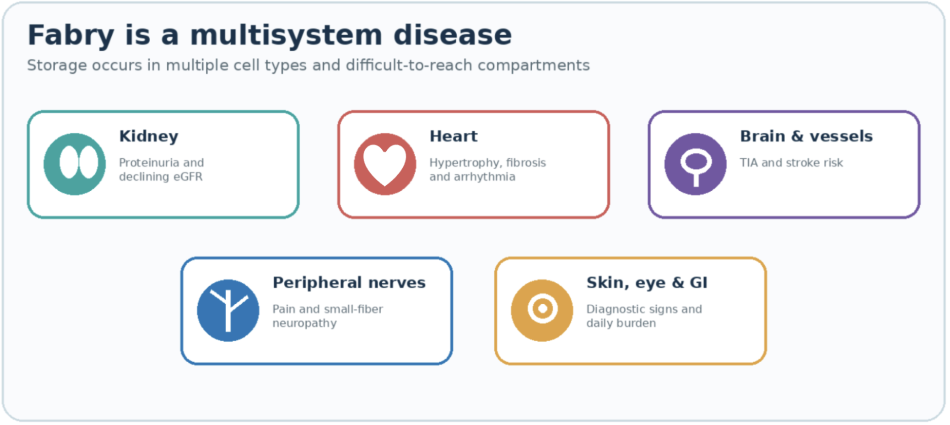
Figure 3. Fabry disease affects multiple organ systems because substrate accumulates across many cell types.
What treatment changes, and what it does not
The unifying goal of every Fabry therapy is to reduce substrate load in the tissues that store it, and the honest way to judge any of them is by two questions: how much substrate does it clear, and can it reach the compartments that matter before those compartments are scarred. The recurring theme across all modalities is that clearing storage early preserves function, while established fibrosis in the heart and kidney is not undone by anything currently available.
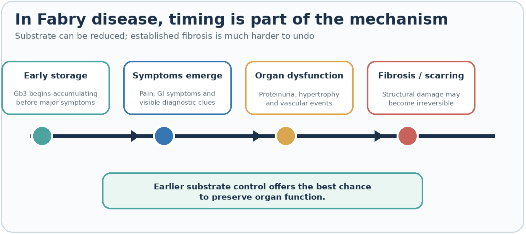
Figure 4. Treatment has the greatest potential before progressive organ injury becomes established fibrosis.
Enzyme replacement therapy
Enzyme replacement therapy, or ERT, has been the backbone of Fabry treatment since 2001 and remains the standard of care. The idea is direct: infuse a recombinant version of alpha-galactosidase A and let it be taken up by cells to clear stored Gb3. Two long-established products, agalsidase beta (Fabrazyme) and agalsidase alfa (Replagal, marketed outside the United States), are given by intravenous infusion every two weeks. A third and newer option, pegunigalsidase alfa (Elfabrio), was approved by the FDA in 2023; it is a PEGylated, plant-cell-produced enzyme engineered for a substantially longer circulating half-life and potentially reduced immunogenicity, though it is still an every-two-week infusion.
Before and after, at the tissue level, ERT is most convincing in the vascular endothelium: biopsy studies show clearance of Gb3 from endothelial cells, and plasma lyso-Gb3 falls with treatment. Renal and cardiac function tend to stabilize, particularly when therapy starts early, and outcomes are meaningfully better than the untreated natural history.
The limits are equally real and should be stated plainly. ERT has a short half-life, so substrate begins re-accumulating between doses, and the every-other-week infusion, often four hours in a chair, is a lifelong commitment that patients describe as a heavy burden. Recombinant enzyme does not reach all compartments equally; podocytes and cardiomyocytes clear less completely than endothelium, and the enzyme does not cross the blood-brain barrier. A meaningful fraction of patients, especially classically affected men, develop anti-drug antibodies that can blunt efficacy (neutralizing anti-drug antibodies have been reported in roughly 40% of classically affected men treated with agalsidase beta; rates are substantially lower in women and in later-onset patients — PMIDs 22227322, 30064518, 30093456). Furthermore, ERT has not been shown to reverse established organ damage: once glomerulosclerosis or myocardial fibrosis is present, the infused enzyme does not clear it. This is why the field keeps returning to the same conclusion, that ERT started before organ damage is a different proposition than ERT started after.

Oral chaperone therapy: migalastat (Galafold)
Migalastat, sold as Galafold, takes a different route. It is a small oral molecule, a pharmacological chaperone that binds the patient's own mutant enzyme, stabilizes its folding, and helps traffic it to the lysosome where it can work. Approved in 2018 in the United States, it replaces biweekly infusions with a pill taken every other day.
The decisive caveat is that migalastat only works if the patient's specific GLA mutation produces an enzyme the chaperone can actually stabilize, an "amenable" variant. Amenability is determined by a standardized laboratory assay, and depending on the population and the assay, roughly 35% to 50% of catalogued GLA variants are amenable; the share of *patients* who are amenable differs, because variant frequencies are not equal. For the majority of patients whose variants are not amenable, the drug is simply not an option. For those who are amenable, migalastat lowers lyso-Gb3, has shown reductions in left ventricular mass, and stabilizes kidney function, with the considerable quality-of-life advantage of oral, non-immunogenic dosing. It is a genuine advance for a defined subset, but only for that subset.
Substrate reduction therapy
Substrate reduction therapy, or SRT, attacks the problem from the opposite end. Rather than replacing the missing enzyme or stabilizing a mutant one, SRT uses an oral inhibitor of glucosylceramide synthase to slow the upstream synthesis of the glycosphingolipids that accumulate. Its conceptual appeal is that it is genotype-independent, so it would apply regardless of variant, and that certain candidates are designed to cross into the central nervous system.
Two agents have been furthest along in Fabry, and their track record is a useful reality check. Lucerastat completed a pivotal Phase 3 program that did not meet its primary endpoint. Venglustat, developed by Sanofi, missed the primary endpoints of its Phase 3 Fabry program in a readout reported in early 2026, even as the same molecule succeeded in a Phase 3 study of type 3 Gaucher disease. As of now, no substrate reduction therapy is approved for Fabry disease. The class remains a scientifically reasonable idea that has not yet delivered a positive pivotal Fabry result, and it is worth watching without being counted on.
Gene and cell-based therapy
The newest wave aims to give patients a durable internal source of the enzyme rather than a repeated external one. Two broad strategies are in development. In vivo AAV approaches deliver a functional GLA gene using an adeno-associated virus vector, typically targeting the liver as an enzyme factory. Ex vivo approaches engineer the patient's own cells, usually hematopoietic stem or progenitor cells, with a lentiviral vector so that the modified cells and their progeny secrete alpha-galactosidase A, which neighboring cells then take up through the mannose-6-phosphate pathway, the same cross-correction biology that makes ERT possible in the first place.
The promise is durability: sustained enzyme production and lower substrate from a single course, with the potential to reach compartments that biweekly infusions serve poorly. The longest running AAV trial is with Sangamo, an AAV6-based viral therapy for Fabry. As of the most recently reported public results of the STAAR study, patients followed out to approximately 4.5 years had not resumed ERT. However, even in Sangamo's own ST-920 data, a subset of Fabry patients showed alpha-galactosidase A activity waning from peak toward the normal range over time, which is consistent with the early and progressive expression decline seen across liver-directed AAV gene therapies, a limitation compounded because anti-capsid immunity makes AAV a one-time, non-repeatable treatment.
The durability of ex vivo lentiviral approaches is also an open question, and is influenced by conditioning intensity, since the regimen used to make room for engineered cells can range from heavy myeloablative chemotherapy to far gentler reduced-intensity protocols. The choice between reduced-intensity conditioning (RIC) and fully myeloablative conditioning (MAC) has large consequences for toxicity, patient eligibility and overall enthusiasm for administering the therapy. For instance, a Fabry therapy conducted in Canada under the ongoing FACTS trial (NCT02800070) has published follow-up out to 5 years in 5 patients (PMID 39794302). In that cohort, reduced-intensity conditioning with low-dose melphalan was reported to be well tolerated, engraftment persisted over years of follow-up, and 3 of the 5 patients elected to stop ERT. At year 4, one of the three went back on ERT at the recommendation of a new attending physician as a precaution. AVROBIO in-licensed the approach and carried it into IND-enabling work with the FDA. Three additional patients were treated using the same reduced-intensity conditioning with low-dose melphalan, with reported engraftment and lyso-Gb3 control similar to the FACTS cohort. AVROBIO then treated one patient with high-dose busulfan for fully myeloablative conditioning; their investor materials reported a higher level of engraftment. When AVROBIO extended the trial to 5 more patients using high-dose busulfan, the protocol had also changed in other respects: cell manufacturing was modified and the washout step was extended from 24 to 72 hours. No detectable engraftment was observed in that cohort. AVROBIO subsequently decided to stop further development of their Fabry program. Glafabra Therapeutics, where the author works, is developing this approach with a later-generation lentiviral vector and intends to seek an IND with the FDA. Glafabra’s current plan is to use reduced-intensity conditioning, consistent with the regimen used in the published FACTS cohort. Glafabra’s product candidate is investigational. It has not been evaluated in any clinical trial conducted by Glafabra and has not been approved by the FDA or any other regulator; whether the approach is safe or effective in Fabry patients is the question its planned trials are designed to answer. There is no assurance that the candidate will advance into clinical trials or reach patients.
Antibody reactions are an important confounder that can alter the effectiveness of all therapies. The immune system can be induced or primed to interfere with therapeutic response. Earlier in this report, immune reactions to ERT were described, affecting roughly 40% of classically affected men treated with agalsidase beta. A second form of antibody response affects the delivery of AAV therapies. Pre-existing neutralizing antibodies to the AAV capsid are a standard exclusion criterion in AAV trials. Reported seroprevalence varies by capsid serotype, assay, and geography; across studies a substantial share of adults — for some serotypes and populations on the order of 40% — would be excluded on this basis (PMIDs 20095819, 35156839, 38323309). Further once a patient has been administered an AAV therapy, they cannot be administered another dose, because the initial administration triggers a strong anti-capsid antibody response that makes re-administration unsafe. The bottom line for the new gene therapies: durability, redosability, and conditioning burden are the three axes on which these programs will be judged.
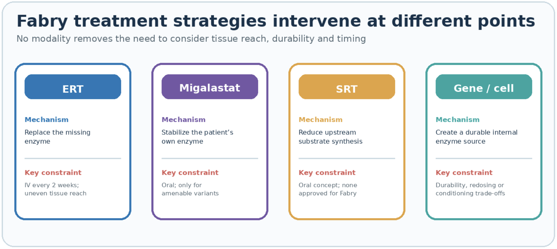
Figure 5. Current and emerging Fabry therapies intervene at different points in the disease pathway.
The through-line
Fabry is a lifelong accumulation disease that begins early in life; symptoms often appear in childhood, before damage to the kidney and heart is established. Every therapy on the market, or in development, focuses on lowering substrate in the tissues that store the build up of toxic lipid. The difference between a good and a poor outcome is usually a difference in timing rather than in the choice of mechanism. ERT stabilizes and is the proven standard of care, but it demands lifelong infusions and does not reverse fibrosis. Migalastat offers oral convenience to the subset with amenable variants; substrate reduction remains unproven in Fabry specifically. Gene and cell-based approaches are chasing the durability that the others lack, and will be measured on how completely and durably they can clear substrate before the organs scar. The biology has been understood for decades. The unfinished work is reaching every compartment, early enough, and keeping it cleared.

Clinical background drawn from Fabry Disease, GeneReviews and StatPearls (NCBI Bookshelf); therapy landscape from recent peer-reviewed reviews of Fabry therapeutics and "New Drugs Available for Fabry Disease" (Kidney and Blood Pressure Research, Karger). Pegunigalsidase alfa (Elfabrio) FDA approval, 2023. Migalastat amenability per the standardized GLP-HEK assay. Lucerastat Phase 3 results per Nature Communications (2025); venglustat Phase 3 Fabry outcome per company disclosures reported in early 2026. FACTS study data per Nature Communications 2021 (PMID 33633114) and Clinical and Translational Medicine 2025 (PMID 39794302).
Register for FREE to comment or continue reading this article. Already registered? Login here.
0